

How to Choose an Ophthalmic Diagnostic Setup for an Eye Clinic
The right ophthalmic diagnostic setup is the one that matches the patients you actually see, the space you actually have, and the services you plan to offer over the next five years. Buying the most advanced device on the market rarely makes a clinic better. Buying the right combination of devices, in the right order, almost always does.
At Medeon Med, we work with eye clinics at every stage, from single-room practices opening their doors to multi-specialty eye hospitals adding retina and glaucoma services. This guide walks through the decisions that matter most when you plan your diagnostic setup.
Define Your Eye Clinic’s Clinical Scope Before Choosing an Ophthalmic Diagnostic Setup
Before comparing brands or specifications, write down what your clinic will do. A general ophthalmology practice that handles refraction, cataract screening, and routine follow-ups needs a very different setup from a clinic focused on diabetic retinopathy or glaucoma management.
Ask these questions first:
- What share of your patients come for refraction and spectacle prescriptions?
- Will you perform cataract or refractive surgery, and do you need pre-operative biometry?
- Do you expect a high volume of diabetic or hypertensive patients needing retinal screening?
- Will you manage glaucoma patients long term, with regular visual field and nerve fibre assessments?
- Do you plan to see children, who need faster and more patient-friendly testing?
Your answers define which devices are essential on day one and which can wait until patient volume justifies them.
Core Ophthalmic Diagnostic Equipment Every Eye Clinic Needs
Almost every eye clinic, regardless of specialty, relies on a basic group of diagnostic tools.
Auto refractometer or auto refkeratometer
This gives a fast, objective starting point for refraction and corneal curvature. A combined refkeratometer saves space and is useful if you plan contact lens fitting or cataract work.
Phoropter or trial lens set
Subjective refraction still depends on the clinician. A manual phoropter is reliable and affordable, while a digital phoropter speeds up high-volume clinics and reduces examiner fatigue.
Visual acuity chart
Digital LCD charts are now common because they offer randomised optotypes, contrast testing, and paediatric options in a compact space.
Slit lamp
This is the heart of the eye examination. Choose a model with good optics, stable illumination, and the option to add a camera module later for documentation and teaching.
Tonometer
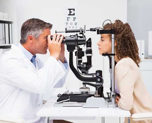
Measuring intraocular pressure is essential for glaucoma screening. Non-contact tonometers are quick and need no anaesthetic drops, which suits busy screening clinics. Applanation tonometers mounted on the slit lamp remain the clinical reference for diagnosis and follow-up.
Ophthalmoscope
Direct and indirect ophthalmoscopes allow fundus examination. Indirect ophthalmoscopy is important for a full peripheral retinal view.
Advanced Ophthalmic Diagnostics: When and Why to Add Them
Once the basics are in place, advanced imaging and testing devices expand what your clinic can diagnose in-house instead of referring out.
Fundus camera
A fundus camera records the retina for documentation, comparison over time, and patient education. Non-mydriatic models capture images without dilation, which is valuable for diabetic screening programmes.
Optical Coherence Tomography (OCT)
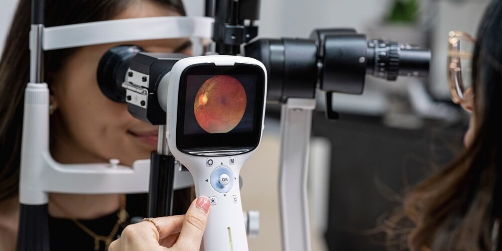
OCT produces cross-sectional images of the retina and optic nerve. It has become central to managing macular disease, diabetic macular oedema, and glaucoma. If your clinic sees many retinal or glaucoma patients, OCT is often the single most valuable advanced investment.
Automated perimeter (visual field analyser)
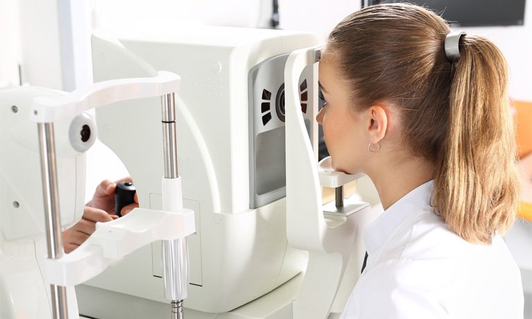
Glaucoma and neuro-ophthalmic conditions require reliable visual field testing. Look for standard testing strategies, a good normative database, and software that tracks progression across visits.
Optical biometer and A-scan
For cataract surgery, accurate axial length and keratometry measurements drive intraocular lens calculations. Optical biometers are fast and non-contact; ultrasound A-scan remains necessary for dense cataracts where optical methods fail. Cataract surgery also relies on an ophthalmic microscope for clear, magnified visualization.
Corneal topographer
Essential for refractive surgery screening, keratoconus detection, and specialty contact lens fitting.
B-scan ultrasound
When the media is opaque, for example with dense cataract or vitreous haemorrhage, B-scan lets you assess the posterior segment.
Plan Your Eye Clinic Workflow for Ophthalmic Diagnostic Equipment in Dubai
A well-chosen device in the wrong place slows the entire clinic. Map the patient journey from registration to final consultation.
A common flow places the auto refractometer and non-contact tonometer at a pre-testing station handled by a technician. Patients then move to the refraction lane and finally to the doctor’s slit lamp room. Imaging devices such as OCT and fundus cameras often sit together in a dedicated diagnostic room, so a single trained technician can operate them.
Consider these practical points:
Room size and lighting
Refraction lanes need the correct testing distance, or a mirror arrangement for shorter rooms. Imaging rooms need controllable lighting.
Combined workstations
Motorised instrument tables and combo units that hold a slit lamp and phoropter save floor space in smaller clinics.
Patient flow
Avoid bottlenecks by not placing all high-demand tests in one room.
Ophthalmic Diagnostic Setup Connectivity and Data Management
Modern ophthalmology runs on images and data. Devices that connect to your electronic medical records or a central image management system save time and reduce transcription errors.
When evaluating equipment, ask whether it supports standard data export formats such as DICOM, whether it can connect to your existing software, and how patient data is stored and backed up. Being able to compare OCT scans or visual fields over several years is clinically important, so data continuity matters as much as image quality.
Evaluate the Total Cost of Your Ophthalmic Diagnostic Setup
The purchase price is only part of the cost. Before you decide, consider:
- Service and maintenance: Is local technical support available, and how quickly can an engineer reach your clinic?
- Spare parts and consumables: Printer paper, chin rest papers, bulbs, and probe covers add up over time.
- Software updates and licences: Some imaging devices charge for upgrades or additional analysis modules.
- Training: Staff need proper training to get reliable, repeatable results.
- Warranty terms: Understand what is covered and for how long.
A slightly more expensive device with strong local support often costs less over its lifetime than a cheaper one that sits idle waiting for repairs.
Plan Your Ophthalmic Diagnostic Setup in Phases
Few clinics buy everything at once. A phased approach protects cash flow while still building a complete setup.
- Phase one: refraction equipment, slit lamp, tonometer, ophthalmoscopes, and visual acuity chart.
- Phase two: fundus camera and biometry if you begin cataract surgery.
- Phase three: OCT, perimeter, and topography as specialty services grow.
When planning phase one, leave space, power points, and network connections for future devices.
Why Work With Medeon Med for Ophthalmic Diagnostic Equipment in Dubai
Choosing ophthalmic diagnostic equipment is a long-term decision. Medeon Med helps eye clinics assess their clinical scope, plan room layouts, compare devices across budgets, and arrange installation, training, and after-sales service. Whether you are opening a new practice or upgrading an established one, our team can help you build a diagnostic setup that fits your patients and grows with your clinic.
Ready to plan your eye clinic setup? Contact Medeon Med today for a consultation and a tailored equipment proposal.